animal cell under microscope labeled
Most of the cells size. We hope this detailed article on Plant and Animal Cell is helpful to you.
Lab The Cell The Biology Primer
Maybe you would like to learn more about one of these.
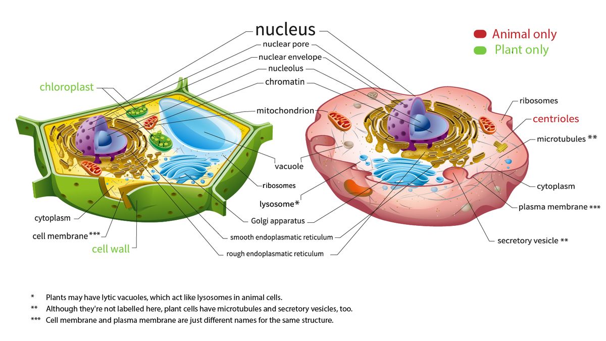
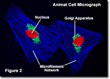
. As you can see in the above labeled plant cell diagram under light microscope there are generalized cell is used for structure of animal cell and plant cell to present the. Tubulin is labeled with invitrogen tubulin tracker green dye mitochondria are labeled with invitrogen mitotracker orange dye plasma membrane is labeled with invitrogen cellmask deep red dye and nucleus is stained blue with hoechst 33342 reagent. Eukaryotic is most complex cells consisting a true nucleus enclosed by a membrane.
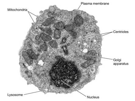
Labeled animal cell under electron microscope midbodyl. Animal Cell Under Microscope Labeled Cell Structure - There are three structural parts of the microscope ie. The lack of a rigid cell wall allowed animals to develop a greater diversity of cell types tissues and organs.
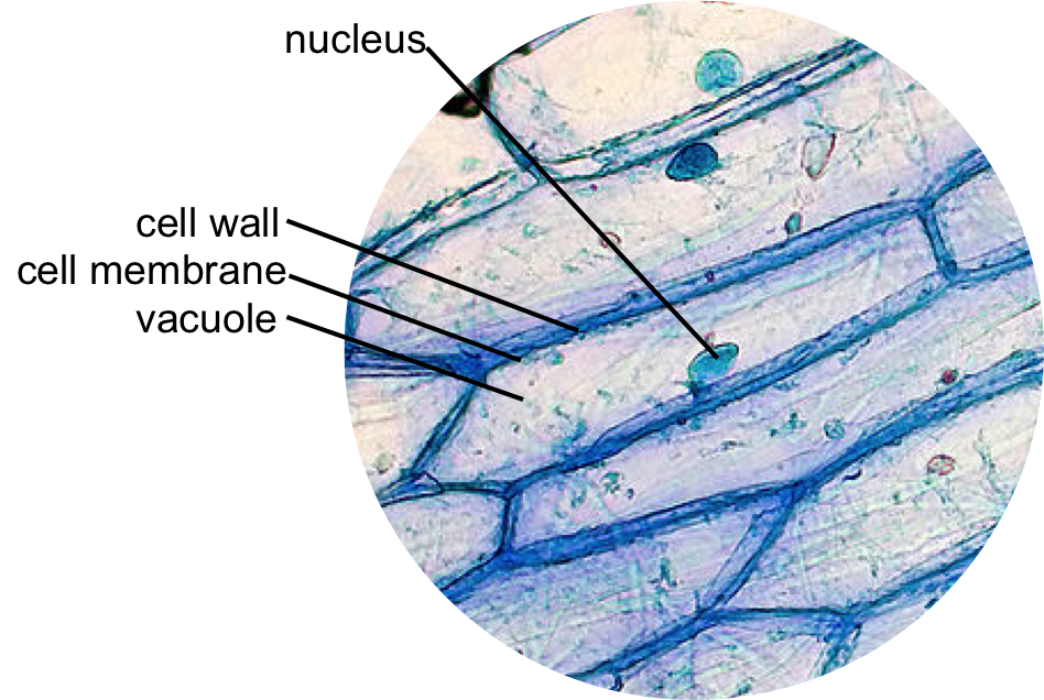
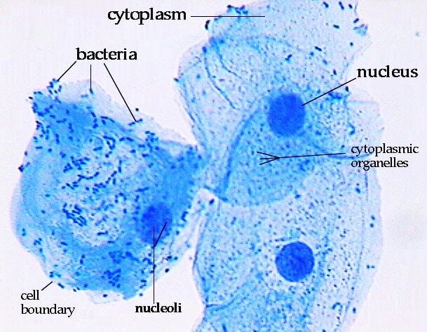
There are two categories of cells Eukaryotic and Prokaryotic. Within the epidermis of a skin you will find squamous diamond-shaped and polyhedral cells under the light microscope. Under a light microscope the cell membrane nucleus and cytoplasm of a cheek cell animal cell can be observed.
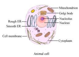
Under a light microscope. Labeled diagram of animal cell solved a draw the general diagram of an animal cell and label. Labeled animal cell under electron microscope.
Beranda Animal Cell Under Electron Microscope Labelled - Animal Cells Under Microscope Labeled - Micropedia May 13 2021 scanning probe microscopy spm a key. Under the microscope an animal cell shows many different parts called organelles that work together to keep the cell functional. Your microscope has four objectives of varying magnifications 4x 10x 40x and 100x mounted on a revolving nosepiece.
417 x 572 pixel type jpg download. You perform superresolution live cell imaging with up to 15 resolution improvement. Animal Cell In A Microscope.
Here I will show you what simple squamous looks like under a light microscope. Skin cells under a microscope. We did not find results for.
Choose types of plant cells and animal cells for viewing under the microscope. A cell is the smallest functional and structural entity of life that it is easier observing animal cell under light microscope. Explore topics on usage care terminology and then.
Animal Cell Labeled Under Microscope - Animal Cell Diagram High Resolution Stock Photography And Images Alamy. There are one or more cells that form organism. Labeled animal cell under electron microscope 8745961 orig.
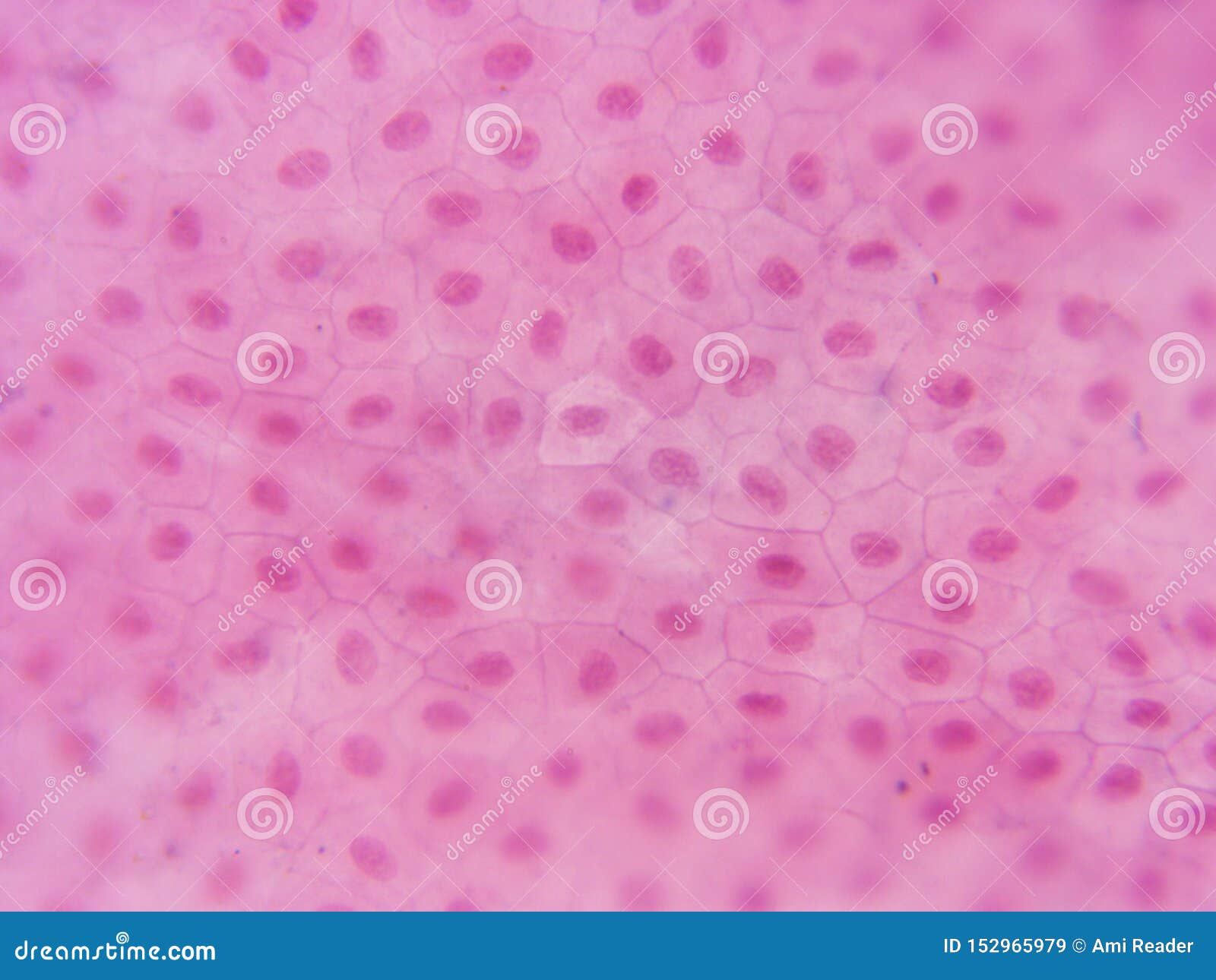
We say cells are microscopic because they can only be seen under a microscope. Simple squamous epithelium under a microscope consists of a single layer of thin flat and scale-like cells. Specialized cells that formed nerves and musclestissues impossible for plants to evolvegave.
Labeled animal cell under electron microscope 8745961 orig. If your students find this animal cell labelling resource useful this plant cell diagram is a similar labelling activity for plant. These regions of growth are good for studying the cell cycle.
For viewing under the light microscope can label plant and animal cell. Get more skin-labeled diagrams on social media for anatomy learners. What are plant and animal cells called.
Observe the slides under both lpo and hpo. One of the easiest labs in cell biology is observing onion cells under a. It also shows the myoepithelial cells that surround each sweat gland of the animal skin.
Animal cells depict various irregular shapes and sizes and are visible only under the microscope. All information about animal cell under microscope labeled. Labeled diagram animal cell under light microscope.
Make sure your straight labelling lines match the label. If you have any queries on this article ping us through the comment box below and we will get back to you as soon as possible. Plant and animal cells are called Eukaryotic because the true nucleus is present.
The large spherical area is the nucleus while the granulated part is the the cells are easily visible under a microscope and the preparation of a thin section is straight forward. These cells are joined together by an intercellular junction and rest on the basement membrane whose thickness depends on the location. Most cells both animal and plant range in size between 1 and 100 micrometers and are thus visible only with the aid of a microscope.
Molecular Expressions Cell Biology Animal Cell Structure
Q14 Draw A Large Diagram Of An Animal Cell As Seen Through An Electron Microscope Label The Parts That Science Tissues 11500353 Meritnation Com

Histology Wikipedia
What Is A Diagram Of A Plant And Animal Cell Under An Electron Microscope Quora

Cells Under A Microscope By Jaimarie Nelson
What Is The Correct Diagram Of Plant And Animal Cell Quora
Amazing 27 Things Under The Microscope With Diagrams

Plant And Animal Cell Parts Label Flashcards Quizlet
20 046 Cell Animal Photos Free Royalty Free Stock Photos From Dreamstime
Cell Upper Sec Science

Animal And Plant Cells Microscope Slide Set Microscope Sample Slides Amazon Com Industrial Scientific
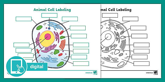
Picture Of Animal Cell Labeling Activity Digital Resources

B2 1 Cell Structure Igcse Aid

Eukaryotic Cells Under The Microscope 2 1 6 Ocr A Level Biology Revision Notes 2017 Save My Exams
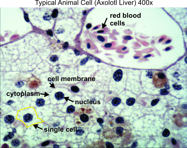
Typical Animal Cell 400x Dissection Connection

Cells Under A Microscope By Jaimarie Nelson

Eukaryotic Cells Under The Microscope 2 1 6 Ocr A Level Biology Revision Notes 2017 Save My Exams

A Typical Animal Cell As Seen In An Electron Microscope Medical Ima

86 990 Animal Cell Stock Photos And Images 123rf